Go beyond western blot images and use the versatile Amersham ImageQuant™ 800 CCD imagers to capture brilliant images to support your scientific research.
Color patterns and color contrasts are the most recognizable way for flowers to attract pollinators like birds and bees. In addition, certain areas of the flower including some pollen and nectar are fluorescent as well, which could potentially aid in the pollination of flowers (1-6). Using multiplex fluorescence imaging we found intricate and detailed fluorescence patterns in flowers which were not detected by visual color inspection. In particular, the central parts exhibit strikingly different fluorescence properties when illuminated with Ultraviolet (UV), visible, and Infrared (IR) light. The potential role of fluorescence in flower signaling and communication is an exciting new field of research [1-6] which requires state of the art imagers. The Amersham ImageQuant™ 800 is an ideal companion with its wide range of narrow range light-emitting diodes (LED) and multiple emission filters, which can be combined freely. The illumination range spans from UV to IR.
The following flowers were imaged using Amersham ImageQuant™ 800 and images were analyzed using ImageQuant™ TL analysis software.
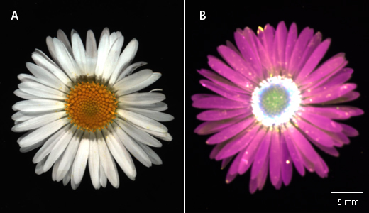
Fig 1. A daisy (Bellis perennis) was imaged with ImageQuant™ 800 using (A) colorimetric and (B) fluorescent imaging modes at UV (magenta), Cy™2 (yellow), Cy™3 (green), and IR long (blue).

Fig 2. A siberian squill (Scilla siberica) was imaged with ImageQuant™ 800 using (A) colorimetric and (B) fluorescent imaging modes at UV (red), Cy™3 (yellow), and Cy™5 (green).
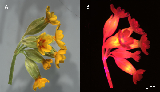
Fig 3. A cowslip (Primula veris) was imaged with ImageQuant™ 800 using (A) colorimetric and (B) fluorescent imaging at Cy™2 (red), Cy™3 (magenta), and IR long (yellow).
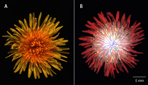
Fig 4. A dandelion (Taraxacum officinale) was imaged with ImageQuant™ 800 using (A) colorimetric and (B) fluorescent imaging at UV (cyan), Cy™2 (red), Cy™3 (yellow), and IR long (blue).
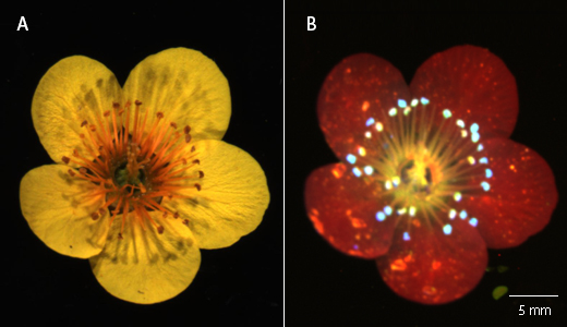
Fig 5. A yellow wood anemone (Anemone ranunculoides) was imaged with ImageQuant™ 800 using (A) colorimetric and (B) fluorescent imaging modes at UV (red), Cy™3 (green), Cy™5 (yellow), and IR long (blue).
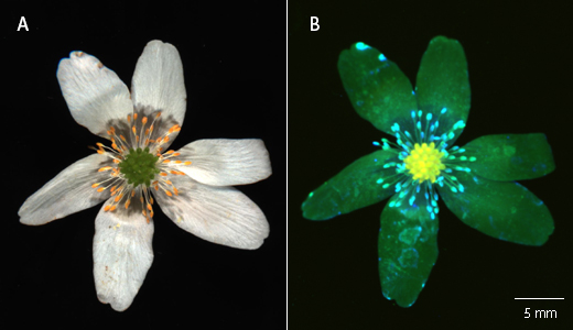
Fig 6. A wood anemone (Anemone nemorosa) was imaged with ImageQuant™ 800 using (A) colorimetric and (B) fluorescent imaging at UV (green), Cy™3 (blue), and IR short (yellow).
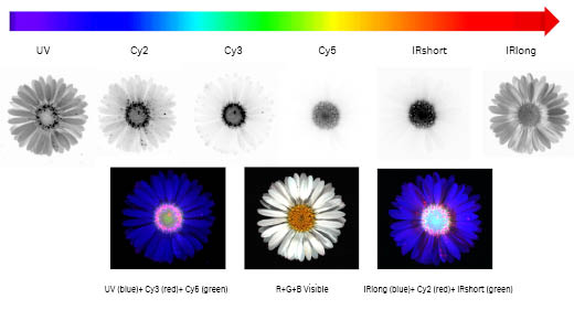
Fig 7. The Amersham ImageQuant™ 800 imager allows high resolution imaging across the entire visible spectrum, from UV to infrared. The daisy Bellis Perennis contains multiple yellow disc florets and white ray florets. The open disc florets display a characteristic fluorescence emission observed with UV, Cy™2, and Cy™3 LED-filter settings compared to the budding florets in the center. In the fake color overlays, these open florets appear as pink dots, approximately 0.5 mm wide. The Amersham ImageQuant™ 800 1×1 binning allows such sub-mm details to be clearly resolved.
References
- O. Ostroverkhova, G. Galindo, C. Lande, J. Kirby, M. Scherr, G. Hoffman and S. Rao, "Understanding innate preferences of wild bee species: responses to wavelength-dependent selective excitation of blue and green photoreceptor types," Journal of Comparative Physiology A, vol. 204, p. 667–675, 2018.
- F. Gandía-Herrero, F. García-Carmona and J. Escribano, "Floral fluorescence effect.," Nature 437, p. 334, 2005.
- C. Van der kooi, A. Dyer, P. Kevan and K. Lunau, "Functional significance of the optical properties of flowers for visual signalling.," Annals of Botany 123, p. 263–276, 2019.
- A. Iriel and M. Lagorio, "Is the flower fluorescence relevant in biocommunication?," Naturwissenschaften 97, p. 915–924, 2010.
- K. Lunau, Z. Ren, X. Fan, J. Trunschke, G. Pyke and H. Wang, "Nectar mimicry: a new phenomenon," Scientific Reports, p. 7039, 2020.
- S. Mori, H. Fukui, M. Oishi, M. Sakuma, M. Kawakami, J. Tsukioka, K. Goto and N. Hirai, "Biocommunication between Plants and Pollinating Insects through Fluorescence of Pollen and Anthers," Journal of Chemical Ecology, vol. 44, p. 591–600, 2018.